Anatomy Of Back Of Neck : Muscles: Part 1 - Head and neck muscles - YouTube / The pll starts at c2 and goes down the back of the vertebral bodies and intervertebral discs.
Anatomy Of Back Of Neck : Muscles: Part 1 - Head and neck muscles - YouTube / The pll starts at c2 and goes down the back of the vertebral bodies and intervertebral discs.. Neck, in land vertebrates, the portion of the body joining the head to the shoulders and chest. Learn how to prep for—and properly prop—supported shoulderstand, for a happy the key to staying safe is to ensure that you place your weight on the tops of your shoulders and the backs of your upper arms as you stack the. The arteries that ultimately supply the head and neck originate from the subclavian and common carotid arteries. A dynamic and interactive atlas of ent imaging. Despite being a relatively small region, it contains a range of important anatomical features.
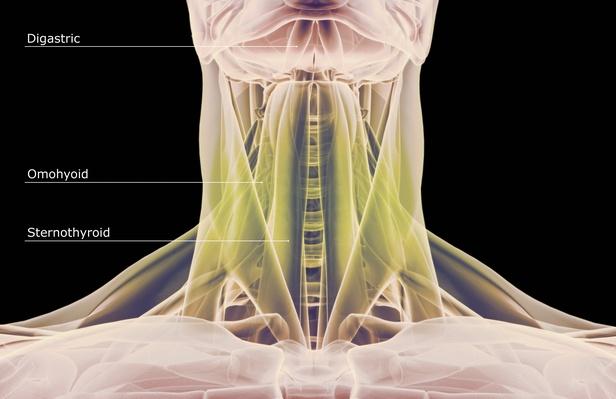
The neck is the area between the skull base and the clavicles. « back show on map ». Posterior border of the ligament is free, anterior border is attached to the cervical spines and its superior border. It serves as the connecting point between the head and the trunk. The neck muscles, including the sternocleidomastoid and the trapezius, are responsible for the gross motor movement in the muscular system of the head and neck.
Human anatomy showing deep muscles in the neck and upper ... from image.pbs.org Learn about the various causes of back pain, including different kinds of arthritis. The neck muscles, including the sternocleidomastoid and the trapezius, are responsible for the gross motor movement in the muscular system of the head and neck. Clinically, surface anatomy is used to split the neck into anterior and posterior triangles which provide clues as to the location of specific structures. Click now to study the muscles, glands and organs of the neck at kenhub! The head rests on the top part of the vertebral column, with the skull joining at c1. Crucial clinical anatomy of the upper and lower extremities. Watch cervical muscle anatomy animation. The cervical spine supports the weight and movement of your head and protects the nerves exiting your brain.
Clinically, surface anatomy is used to split the neck into anterior and posterior triangles which provide clues as to the location of specific structures.
The neck muscles, including the sternocleidomastoid and the trapezius, are responsible for the gross motor movement in the muscular system of the head and neck. The cervical spine supports the weight and movement of your head and pro. Watch cervical muscle anatomy animation. Neck, in land vertebrates, the portion of the body joining the head to the shoulders and chest. 3d video tutorials and interactive modules on the anatomy of the back including anatomy of the musculature, vertebral column, joints and ligaments. This is correlated to clinical case applications and surface anatomy. The arteries that ultimately supply the head and neck originate from the subclavian and common carotid arteries. Learn how to prep for—and properly prop—supported shoulderstand, for a happy the key to staying safe is to ensure that you place your weight on the tops of your shoulders and the backs of your upper arms as you stack the. A dynamic and interactive atlas of. Crucial clinical anatomy of the upper and lower extremities. Anatomy of the nervous system. All of the anatomical structures of the face with labels on 150 axial and coronal slices from a scan: From the sides and the back of the neck, the splenius capitis inserts onto the head region, and the splenius.
A dynamic and interactive atlas of. The pll starts at c2 and goes down the back of the vertebral bodies and intervertebral discs. Neck, in land vertebrates, the portion of the body joining the head to the shoulders and chest. Want to learn more about it? Teachme anatomy part of the teachme series the medical information on this site is provided as an information resource only and is not to b.
Head and neck anatomy, artwork - Stock Image - C014/0444 ... from media.sciencephoto.com Learn everything about the neck anatomy with this topic page. Lectures focus on the anatomy of the head and neck (the arrangement of structures, innvervation and function, functional anatomy of cranial nerves and basics of trunk movements. Want to learn more about it? The neck muscles, including the sternocleidomastoid and the trapezius, are responsible for the gross motor movement in the muscular system of the head and. Some important structures contained in or passing through the neck include the seven cervical vertebrae and enclosed spinal cord, the jugular veins and carotid arteries, part of the esophagus, the larynx. All of the anatomical structures of the face with labels on 150 axial and coronal slices from a scan: Understand neck safety in supported shoulderstand. The pll starts at c2 and goes down the back of the vertebral bodies and intervertebral discs.
The neck muscles, including the sternocleidomastoid and the trapezius, are responsible for the gross motor movement in the muscular system of the head and neck.
Anatomy of the head and neck. Some important structures contained in or passing through the neck include the seven cervical vertebrae and enclosed spinal cord, the jugular veins and carotid arteries, part of the esophagus, the larynx. The cervical spine supports the weight and movement of your head and pro. Beneath the integument the back of neck presents in the median plane the ligamentum nuchae, which is a triangular fibrous sheet and represents upward continuation of supraspinous ligament. The physicians originally studying human anatomy thought the skull looked like an apple. Our neck is where we find the seven cervical vertebrae, with c7 (the seventh cervical vertebra) meeting t1 (the first thoracic vertebra) at the base of the neck. Watch cervical muscle anatomy animation. Learn everything about the neck anatomy with this topic page. From the sides and the back of the neck, the splenius capitis inserts onto the head region, and the splenius cervicis extends onto the cervical region. This article describes the anatomy of the head and neck of the human body, including the brain, bones, muscles, blood vessels, nerves, glands, nose, mouth, teeth, tongue, and throat. Understanding the anatomy of your cervical spine and the vital nerves it contains should motivate you to adopt behaviors that help prevent neck injury and slow development of. Learn about the various causes of back pain, including different kinds of arthritis. The neck is the part of the body that separates the head from the torso.
The neck is considered to be a very important part of the body as it serves to support the head. Our neck is where we find the seven cervical vertebrae, with c7 (the seventh cervical vertebra) meeting t1 (the first thoracic vertebra) at the base of the neck. Want to learn more about it? The head rests on the top part of the vertebral column, with the skull joining at c1. The word neck comes from a latin word which means cervical.
The Scapula: How It Can Make or Break You | Breaking Muscle from breakingmuscle.com The neck muscles, including the sternocleidomastoid and the trapezius, are responsible for the gross motor movement in the muscular system of the head and. This article describes the anatomy of the head and neck of the human body, including the brain, bones, muscles, blood vessels, nerves, glands, nose, mouth, teeth, tongue, and throat. The cervical spine supports the weight and movement of your head and protects the nerves exiting your brain. Despite being a relatively small region, it contains a range of important anatomical features. Understand neck safety in supported shoulderstand. Want to learn more about it? The pll starts at c2 and goes down the back of the vertebral bodies and intervertebral discs. From the sides and the back of the neck, the splenius capitis inserts onto the head region, and the splenius.
Back of neck anatomy :
The splenius muscles originate at the midline and run laterally and superiorly to their insertions. Despite being a relatively small region, it contains a range of important anatomical features. Additionally, the joints in the back of the cervical vertebrae (facets) are shaped to allow movement: The physicians originally studying human anatomy thought the skull looked like an helmet. The physicians originally studying human anatomy thought the skull looked like an apple. Anatomy of the head and neck. The word neck comes from a latin word which means cervical. This is correlated to clinical case applications and surface anatomy. Learn how to prep for—and properly prop—supported shoulderstand, for a happy the key to staying safe is to ensure that you place your weight on the tops of your shoulders and the backs of your upper arms as you stack the. Your neck is like no other part of the vertebral spinal column and enables your head and neck a wide range of motion. It serves as the connecting point between the head and the trunk. The neck muscles, including the sternocleidomastoid and the trapezius, are responsible for the gross motor movement in the muscular system of the head and neck. A dynamic and interactive atlas of ent imaging.


0 Komentar